DIP Joint Fusion PDF Evidence
A protected recovery plan after fusion (arthrodesis) of the small fingertip joint nearest the nail, keeping the fingertip splinted while every other joint stays moving and swelling is controlled, then progressively rebuilding pinch and grip once the bone has joined.
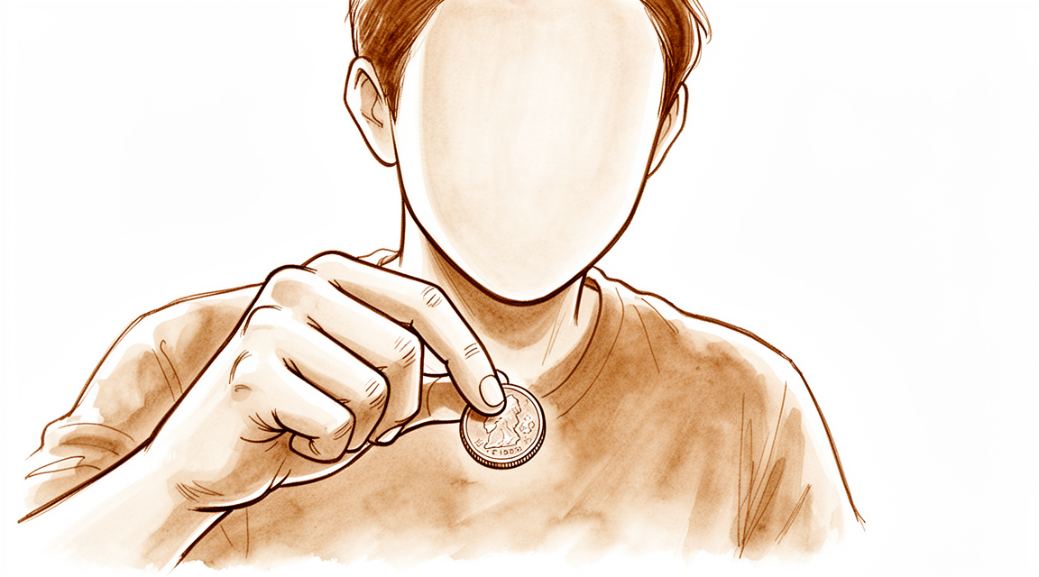
This protocol guides your recovery after a DIP joint fusion (arthrodesis) — an operation that permanently joins the small joint at the very tip of your finger, nearest the nail — with Dr Kieran Hirpara at Mater Private Hospital Rockhampton. It begins with your home exercise program, followed by the structured clinical protocol written for your hand therapist — bring this page or its PDF to your first therapy visit so your rehabilitation stays coordinated. Your therapist may adjust the plan depending on how your recovery progresses.
If you have any concerns about your wound after surgery, get in touch with the rooms. It is often helpful to take a photo of the wound and email it for review.
What to expect
A DIP joint fusion is done when the small joint nearest the nail is worn out and painful — usually from arthritis (the bony lumps called Heberden's nodes), or to remove a troublesome mucous cyst together with the bone spur underneath it. Rather than trying to keep a painful, damaged joint moving, the operation fuses it solid in a slightly bent, functional position (up to about 35°). By design, that joint never moves again — and in exchange the pain goes and the fingertip becomes stable and strong to pinch with. The fixation is usually a small buried headless screw that stays in for good (no need to remove it), or sometimes a K-wire that is taken out at around six weeks. If a mucous cyst was removed, you will also have skin or nail-fold care to do as the area heals.
The whole of your rehabilitation is built around one simple idea: protect the fusion until the bone has joined, but keep everything else moving. The bone typically feels united by about six to eight weeks, with the X-ray catching up by around ten weeks. Until then:
- The fused fingertip is splinted and protected so the healing bone is not disturbed.
- Every other joint keeps moving — the middle joint of the finger, the knuckle, the thumb, the wrist, and all your other fingers — so the hand does not stiffen.
- Swelling is controlled and the scar is managed so the finger stays comfortable and supple.
- Once the bone has joined, pinch and grip are rebuilt gradually rather than all at once.
Precautions and limitations
- Wear your fingertip splint as directed. Early on it is worn continually; later it is worn only for activity. It holds the fused joint still but leaves the middle joint of the finger (the PIP) free to move.
- Do NOT power-grip, pinch hard or lift heavily with the operated finger until the fusion has joined and you are cleared — keep to about 1 kg (≈2 lb) in the first six weeks.
- Keep every other joint moving from the start — the middle and knuckle joints of the finger, the thumb, the wrist, and all your other fingers.
- Keep the dressing dry and the hand elevated in the first 10–14 days to settle swelling, and follow any nail-fold or cyst-site care if a mucous cyst was removed.
- If you have a K-wire, protect it and keep the area clean until it is removed at about six weeks; a buried screw needs no removal.
- Do NOT drive until you are out of the bulky splint and can safely grip and control the wheel — usually around six weeks, at your surgeon's discretion.
For wound, swelling and scar management, see the practice's wound care guidance.
Your exercises
PIP and MCP movement (the joints either side of the fusion)
With your DIP splint on (it holds only the fingertip joint still), bend and straighten the MIDDLE joint of the finger and the big knuckle joint at its base, making a gentle fist and opening fully. The fused tip is not meant to move, but the joints either side of it must stay loose so the finger does not stiffen up. Move smoothly and within comfort.
10 times, 3–4 times a day
Move all your other fingers, thumb and wrist
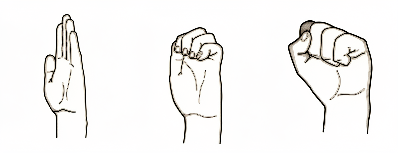
Keep everything that was NOT operated on moving freely from day one: make a full fist and open right out, touch your thumb to each fingertip, and bend your wrist gently up and down. Only the fused fingertip is restricted — the rest of the hand should work as normally as comfort allows so it does not get stiff or weak.
10 of each, several times a day
Kieran Hirpara 4.0
Tendon glides (hook, fist, straight)
Move your fingers through three shapes — a hook (bend the tip and middle joints, knuckles straight), a full fist, then a flat straight hand — pausing briefly in each. This keeps the tendons gliding smoothly through the healing area so they do not stick down. Keep the splinted fingertip comfortable throughout; the glide is for the rest of the finger.
5–10 of each shape, 3 times a day
Swelling control (elevation and compression)
Keep your hand raised above the level of your heart as much as you can in the first couple of weeks — propped on a pillow or cushion. Once the wound allows, a light compression sleeve or self-adherent (Coban) wrap on the finger helps settle swelling. Less swelling means an easier, freer-moving hand.
Elevate often through the day; compression as fitted by your hand therapist
Scar massage
Once the wound is fully healed and your hand therapist gives the go-ahead, rub a little plain moisturiser into the scar with small, firm circles for a minute or two. If a mucous cyst was removed, your therapist will also guide care of the nail fold and surrounding skin. This keeps the scar soft and less sensitive.
1–2 minutes, 2–3 times a day, once the wound is healed
Kieran Hirpara 4.0
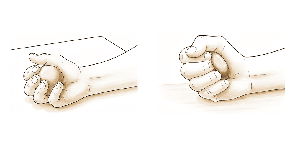
Grip and pinch strengthening (after union)
A LATER exercise — only once the fusion has joined on X-ray and your hand therapist starts it (commonly from around 6–8 weeks). Begin gently squeezing therapy putty and doing light pinches between thumb and fingertip, then build up the effort slowly over the following weeks. Do not power-grip or pinch hard before you are cleared — loading the fingertip too early can disturb the healing bone.
As guided by your hand therapist, building gradually (from ~6–8 weeks)
These are the exercises from your handout. Start them only as guided by Dr Hirpara and your hand therapist, staying within whatever limits you have been given. The early exercises keep the rest of the hand moving freely without disturbing the fused fingertip — movement of the joints either side of the fusion, all your other fingers, thumb and wrist, tendon glides, and swelling control. Grip and pinch strengthening belongs to a later phase and should not be started until the fusion has joined on X-ray and you are specifically cleared. Stop anything that causes sharp pain at the fingertip.
Your clinical protocol
The rest of this page is the staged clinical protocol for rehabilitation after DIP joint (distal interphalangeal) arthrodesis. This section is to be provided to your hand therapist, and each phase opens with a plain-English explanation of what is happening. The principle is protect the arthrodesis site until bony union while preserving full motion at every other joint — the DIP is immobilised in a P2–P3 splint that leaves the PIP free, oedema and scar are managed, and pinch/grip are reloaded progressively only after union.
Prior to treatment, check the patient's operation report and past medical history, and liaise with the treating surgeon regarding the fixation (headless compression screw — buried, no removal — vs K-wire, removed ~6 weeks), the fusion position (slight flexion, up to ~35°), and whether a mucous cyst with skin/nail-fold excision was performed. Clinical union is typically expected around 6–8 weeks with radiographic union around 10 weeks; the rehab timeline below is low-level expert consensus and is subject to surgeon discretion and X-ray confirmation of union before splint weaning and loading.
Phase 1 — protect and settle (weeks 0 to 2)
The first fortnight protects the freshly fixed fusion and settles swelling and the wound, while keeping every uninvolved joint moving so nothing stiffens.
For your hand therapist:
Education and precautions - Bulky surgical dressing/splint with elevation for the first 10–14 days; keep the dressing dry - Protect the arthrodesis site; no loading of the operated fingertip - If a K-wire is present, protect the pin site; review nail-fold/cyst-excision wound where applicable
Management - Wound: surgical dressings as directed; monitor for infection - Oedema: elevation, gentle hand pumping, ice as appropriate - Exercises: AROM of all uninvolved joints — PIP and MCP of the operated finger, thumb, wrist, and all other digits; commence tendon glides as comfort allows
Criteria to progress - Wound settling; swelling controlled; ready to transition into a custom removable DIP-blocking splint at around two weeks
Phase 2 — DIP-blocking splint with activity (weeks 2 to 6)
From about two weeks the bulky dressing is replaced by a custom removable DIP-blocking splint (a Stax/mallet-type orthosis spanning P2–P3) that immobilises only the fingertip joint and leaves the PIP free. It is worn continually through this phase. Full active motion is encouraged everywhere else, swelling and scar are managed, and the fingertip stays unloaded.
For your hand therapist:
Education and precautions - Custom removable DIP-blocking splint (P2–P3, PIP free) worn continually through this phase - No power grasp or pinch; functional load limit ~2 lb (≈1 kg)
Management - Exercises: active PIP, MCP, thumb and wrist motion plus all-other-digit motion; tendon glides (hook, full fist, straight) - Oedema: continue elevation and add compression (Coban/light sleeve) as tolerated - Scar: commence scar massage once the wound is fully healed; nail-fold care where a mucous cyst was excised
Criteria to progress - Maintained PIP/MCP motion; controlled swelling; healed wound; clinical union emerging at around six weeks (proceed to weaning only with X-ray confirmation of union)
Phase 3 — wean the splint and begin gentle strengthening (weeks 6 to 8)
Once the fusion is united on X-ray (clinically ~6–8 weeks), the splint is weaned — worn for activity/protection only — and a K-wire, if used, is removed at about six weeks. Gentle strengthening of pinch and grip begins.
For your hand therapist:
Education and precautions - Wean the DIP splint once union is confirmed — continued protective/activity-only wear as needed; K-wire removed ~6 weeks - Progress loading gradually; functional limit ~5 lb (≈2 kg) from around 8 weeks
Management - Exercises: begin gentle grip and pinch strengthening — therapy putty, light pinch and grip work; continue full motion at all other joints; continue scar management - Reassess any residual swelling or PIP/MCP stiffness and address as needed
Criteria to progress - Radiographic union confirmed; comfortable fingertip; tolerating gentle loading without pain at the fusion site
Phase 4 — progressive strengthening and discharge (weeks 8 to 12)
With the fusion solid, strengthening is progressed towards normal hand function, and restrictions are lifted by around twelve weeks.
For your hand therapist:
Education and precautions - Progressive grip and pinch strengthening; functional limit ~10 lb (≈4.5 kg) at around 10 weeks - No restriction from around 12 weeks, subject to surgeon review
Management - Exercises: graded resistive grip and pinch (putty → grippers → task-specific loading); restore full functional hand use - Consider discharge once a stable, pain-free fingertip with near-normal hand function and strength is achieved - Refer back to the treating surgeon if there is pain over the fusion, concern about union, or a poor functional outcome
Criteria for discharge - United, pain-free fusion; full motion at all non-fused joints; functional pinch and grip restored
Getting back to work and activity
Light everyday use of your other fingers and the rest of the hand is encouraged from the start, within comfort — only the fused fingertip is held back. Driving usually resumes at around six weeks, once you are out of the bulky splint and can grip and control the wheel safely; this is at Dr Hirpara's discretion at your review, so plan for help with transport in the early weeks. Gentle pinch and light grip typically begin around six weeks and build up from about eight weeks, once the bone has joined. Full, heavy or sporting use of the hand is generally reached by around twelve weeks. These timelines are expert-consensus guidelines rather than fixed deadlines — your surgeon's discretion and your X-ray (confirming the bone has joined) come first.
After your protocol
This protocol works alongside the practice's general recovery advice — see managing post-operative pain, wound care and scar management. The phased plan above reflects published rehabilitation guidance after DIP joint arthrodesis, and your ongoing recovery is guided individually by Dr Hirpara and your hand therapist according to how your finger heals.
Evidence & references
DIP Joint Fusion — Procedure Outcomes & Post-operative Rehabilitation (Distal Interphalangeal Arthrodesis)
Topic scope: post-operative rehabilitation after arthrodesis (fusion) of the distal interphalangeal (DIP) joint — most often for end-stage osteoarthritis (Heberden's nodes), or to excise a mucous cyst together with its underlying osteophyte. This is a fusion, not a reconstruction: the joint is deliberately and permanently abolished and set in a slightly flexed, functional position, so the rehabilitation is a protect-to-union pathway built around oedema control, scar/nail-fold management, and preservation of motion at every adjacent joint, followed by progressive reloading — not restoration of DIP motion.
Defining principle of the rehab here: a DIP arthrodesis is meant to stop moving. The single therapeutic goal is to deliver a solid, pain-free, well-aligned bony union while keeping the rest of the hand fully mobile. The fingertip is immobilised in a P2–P3 (Stax/mallet-type) orthosis that blocks the DIP but leaves the PIP free; the deliberate restraints are protection of the fixation and avoidance of pinch/grip loading until union. The principal branch points are the fixation method (buried headless compression screw — no removal — versus K-wire, removed at ~6 weeks) and whether a mucous cyst with skin/nail-fold excision was performed, which adds soft-tissue/scar care. Union, not the calendar, gates splint weaning and loading.
A. PROCEDURE OUTCOMES (fusion union, position, fixation)
DIP arthrodesis is a reliable pain-relieving operation; the principal technical debates are over fixation method and fusion position, not whether to fuse a painful, end-stage joint.
- High union rates with headless compression screw fixation. A series of 64 joints fused with a Herbert-type headless compression screw reported reliable bony union with a low complication profile, supporting the buried-screw construct that requires no later removal [Hand 2010, DOI: 10.1007/s11552-010-9295-3]. Moderate (observational case series).
- Radiographic union averages around ten weeks. A review of DIP arthrodesis techniques reports a mean time to radiographic fusion of approximately 10 weeks, with reported union rates such as ~85% in the Brutus cohort, underlining that clinical comfort precedes full radiographic consolidation [J Hand Surg Am 2013, DOI: 10.1016/j.jhsa.2013.06.010]. Moderate (review of case series).
- Fusion position is a consensus, not a controversy. Technique descriptions place the DIP in slight flexion in a functional position for pinch; dorsal-plate and screw techniques are described with attention to setting and holding this position during fixation [J Hand Surg Am 2018, DOI: 10.1016/j.jhsa.2018.03.049]. Mechanistic / consensus.
- Screw fit depends on bony dimensions. Anatomical sizing work shows the headless screw must be matched to the medullary dimensions of the distal phalanx, informing implant selection and reducing fixation-related complications [Hand 2014, DOI: 10.1007/s11552-014-9679-x]. Mechanistic.
- Fixation choice carries differing complication patterns. Comparative data on K-wire versus headless (Herbert) screw fixation describe differences in infection and hardware-related events, relevant to the protective pin care needed when a K-wire is used and removed at ~6 weeks [J Hand Surg Am 2013, DOI: 10.1016/j.jhsa.2013.01.017]. Moderate (comparative series).
- Acute arthrodesis is an established option in trauma. Primary IP-joint arthrodesis for acute injury is a recognised technique, supporting fusion as a durable solution beyond degenerative disease [J Hand Surg / Thieme 2017, DOI: 10.1055/s-0037-1608691]. Moderate (case series).
B. REHABILITATION / THERAPY EVIDENCE
There are no randomised trials of rehabilitation after DIP arthrodesis. The rehab pathway is built from surgical-outcome timing data (union ~6–8 weeks clinical, ~10 weeks radiographic) plus published hand-therapy protocols and standard hand-therapy practice. The therapeutic logic is to immobilise only the fused joint, keep every other joint moving, control swelling and scar, and reload pinch/grip only after union.
- Immobilise the DIP, free the PIP. Published finger-fusion therapy protocols use a custom removable DIP-blocking (Stax/mallet-type) orthosis spanning P2–P3 that holds the fingertip joint while leaving the PIP free for active motion — continual early wear, weaning to activity-only after X-ray union [TCO; Hand Wisconsin; Alaska Ortho; Melbourne Arm Clinic protocols, URLs below]. Weak (consensus / published protocols).
- Preserve motion at all uninvolved joints from day one. Active motion of the PIP, MCP, thumb, wrist and all other digits, plus tendon glides, is standard hand-therapy practice to prevent stiffness while the DIP consolidates [published protocols, URLs below]. Consensus / standard practice.
- Oedema and scar control are routine adjuncts. Elevation and compression for swelling, and scar massage once healed (with nail-fold care after mucous-cyst excision), follow standard hand-therapy practice rather than trial evidence. Consensus / standard practice.
- Loading is gated by union, not by date. Protocols withhold power grasp/pinch until the fusion is radiographically united, then progress strengthening gradually — reflecting the ~10-week mean radiographic union from the outcome literature [J Hand Surg Am 2013, DOI: 10.1016/j.jhsa.2013.06.010]. Weak–moderate (timing anchored to outcome series; rehab schedule consensus).
Recovery trajectory (expected, evidence-anchored)
| Phase | Window | Restraint | Hand use / therapy focus | Strength / load | Notes |
|---|---|---|---|---|---|
| 1 — Protect & settle | Week 0–2 | Bulky dressing/splint; DIP unloaded | Elevation; AROM of all uninvolved joints (PIP, MCP, thumb, wrist, other digits); begin tendon glides | None to the fingertip | Keep dressing dry; review pin/cyst-excision wound |
| 2 — DIP-blocking splint with activity | Week 2–6 | Custom P2–P3 DIP-block, PIP free, worn continually | Active PIP/MCP/thumb/wrist + all-other-digit motion; tendon glides; oedema (Coban/sleeve); scar massage once healed | No power grasp/pinch; ~2 lb (≈1 kg) limit | Clinical union emerging ~6 wk |
| 3 — Wean splint & gentle strengthening | Week 6–8 | Splint weaned once united on X-ray; K-wire out ~6 wk | Begin gentle grip/pinch (putty, light pinch/grip); continue full motion elsewhere; continue scar care | ~5 lb (≈2 kg) from 8 wk | Buried screw needs no removal |
| 4 — Progressive strengthening & discharge | Week 8–12 | Restrictions lifting | Progressive grip/pinch strengthening; restore full hand use | ~10 lb (≈4.5 kg) at 10 wk; no restriction ~12 wk | Discharge when fusion solid + pain-free |
(Phase windows mirror the precautions and recovery-curve structure in the patient protocol; clinical union ~6–8 weeks and radiographic union ~10 weeks are anchored to the outcome series, while the exact phase timings are low-level expert consensus, not trial-derived deadlines, and are subject to surgeon discretion and X-ray confirmation of union.)
C. KEY CONTROVERSIES / EVIDENCE QUALITY
- Fixation method. Buried headless compression screw (no removal) versus K-wire (removed ~6 weeks) — both achieve union; the comparative literature describes differing infection and hardware-event profiles, and the choice drives whether pin-site protection is needed in rehab [DOI: 10.1007/s11552-010-9295-3; DOI: 10.1016/j.jhsa.2013.01.017]. Moderate.
- Fusion position. Slight flexion (up to ~35°) in a functional pinch position is a settled consensus across technique descriptions, not a live controversy [DOI: 10.1016/j.jhsa.2018.03.049]. Consensus.
- Union timing. Clinical comfort (~6–8 weeks) precedes radiographic union (~10 weeks mean), so splint weaning and loading should follow the X-ray rather than the calendar [DOI: 10.1016/j.jhsa.2013.06.010]. Moderate.
- Rehabilitation schedule. No RCTs exist for DIP-fusion rehab; phase timings are derived from published therapy protocols and standard hand-therapy practice anchored to surgical union data. Low-level expert consensus.
- Mucous-cyst cases. Excision of a mucous cyst with its osteophyte adds skin/nail-fold and scar care to the standard fusion rehab; this is a soft-tissue management addition rather than a change to the bony-union pathway. Consensus / standard practice.
D. EVIDENCE STRENGTH FLAGS (summary)
- MODERATE (observational case series / reviews): reliable bony union with headless compression screw fixation; mean radiographic union ~10 weeks (~85% union, Brutus); differing complication profiles by fixation method; acute IP arthrodesis as an established trauma option.
- CONSENSUS / MECHANISTIC: slight-flexion functional fusion position; screw sizing to phalangeal dimensions; immobilise-the-DIP / free-the-PIP splinting principle.
- WEAK / LOW-LEVEL CONSENSUS: the specific phased rehabilitation schedule (no RCTs; derived from published therapy protocols + standard hand-therapy practice, anchored to union timing); exact phase timings and load limits (typical guides, not trial-derived); oedema/scar adjuncts (standard practice).
CITATIONS
RAG corpus (180,000+ Orthopaedic articles)
- Distal interphalangeal joint arthrodesis using a dorsal plate: technique and fusion position. J Hand Surg Am. 2018. DOI: 10.1016/j.jhsa.2018.03.049
- Distal interphalangeal joint arthrodesis with the Herbert headless compression screw: union and complications in 64 joints. Hand (N Y). 2010. DOI: 10.1007/s11552-010-9295-3
- Distal interphalangeal joint arthrodesis: review of techniques and outcomes (mean ~10-week radiographic fusion; Brutus ~85% union). J Hand Surg Am. 2013. DOI: 10.1016/j.jhsa.2013.06.010
- K-wire versus Herbert screw fixation for distal interphalangeal joint arthrodesis: infection and hardware events. J Hand Surg Am. 2013. DOI: 10.1016/j.jhsa.2013.01.017
- Anatomical sizing of headless compression screws for distal phalangeal fixation. Hand (N Y). 2014. DOI: 10.1007/s11552-014-9679-x
- Acute interphalangeal joint arthrodesis in trauma. J Hand Surg / Thieme. 2017. DOI: 10.1055/s-0037-1608691
DIP-fusion rehabilitation literature (URLs)
- Twin Cities Orthopedics — Distal Interphalangeal (DIP) Joint Fusion post-op protocol. https://www.tcomn.com/wp-content/uploads/2016/06/Distal-Interphalangeal-DIP-Joint-Fusion.pdf
- Hand Wisconsin — Finger-joint fusion therapy protocol. https://handwisconsin.com/wp-content/uploads/2016/09/fusion-finger-joint-therapy-protocol.pdf
- Alaska Orthopaedics — Arthrodesis (DIP / PIP or MCP) joint fusion protocol. https://www.akortho.com/wp-content/uploads/Arthrodesis-DIP-PIP-or-MCP-Joint-Fusion.pdf
- Melbourne Arm Clinic — PIP / DIP arthrodesis rehabilitation protocol. https://melbournearmclinic.com.au/orthopaedic-rehabilitation/shoulder-rehabilitation/pip-dip-arthrodesis-protocol/